 quoneon
A diffusion tensor is a symmetric matrix describing directional diffusion in a voxel. Its eigenvalues summarize diffusion along three principal directions.
Mathematical principle
MD = (λ₁ + λ₂ + λ₃)/3
D is the tensor, g a unit gradient direction, b the diffusion weighting, and S₀ the unweighted signal.
Input definitions
| Item | Meaning |
|---|---|
| Diffusion images | Signal measurements across diffusion directions. |
| Gradients and b-values | Correspond to the image-volume order. |
| Spatial context | Orientation and the analysis mask. |
Analysis choices
| Item | Meaning |
|---|---|
| Tensor fit | Record the estimation procedure. |
| Preprocessing | Record corrections applied to the data. |
| Measurements | Distinguish FA, MD, and directional diffusivities. |
Diffusion imaging reference views
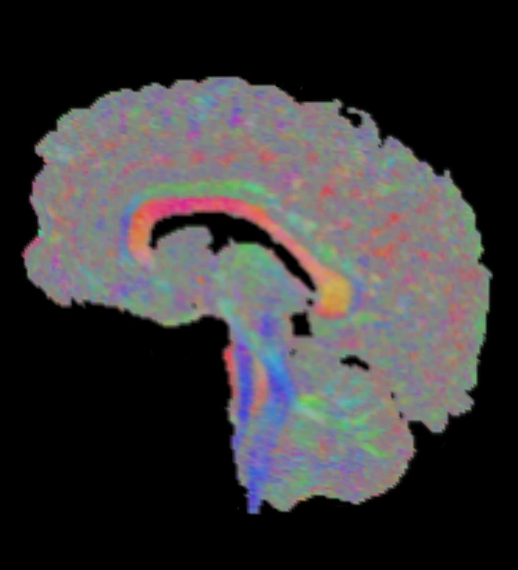
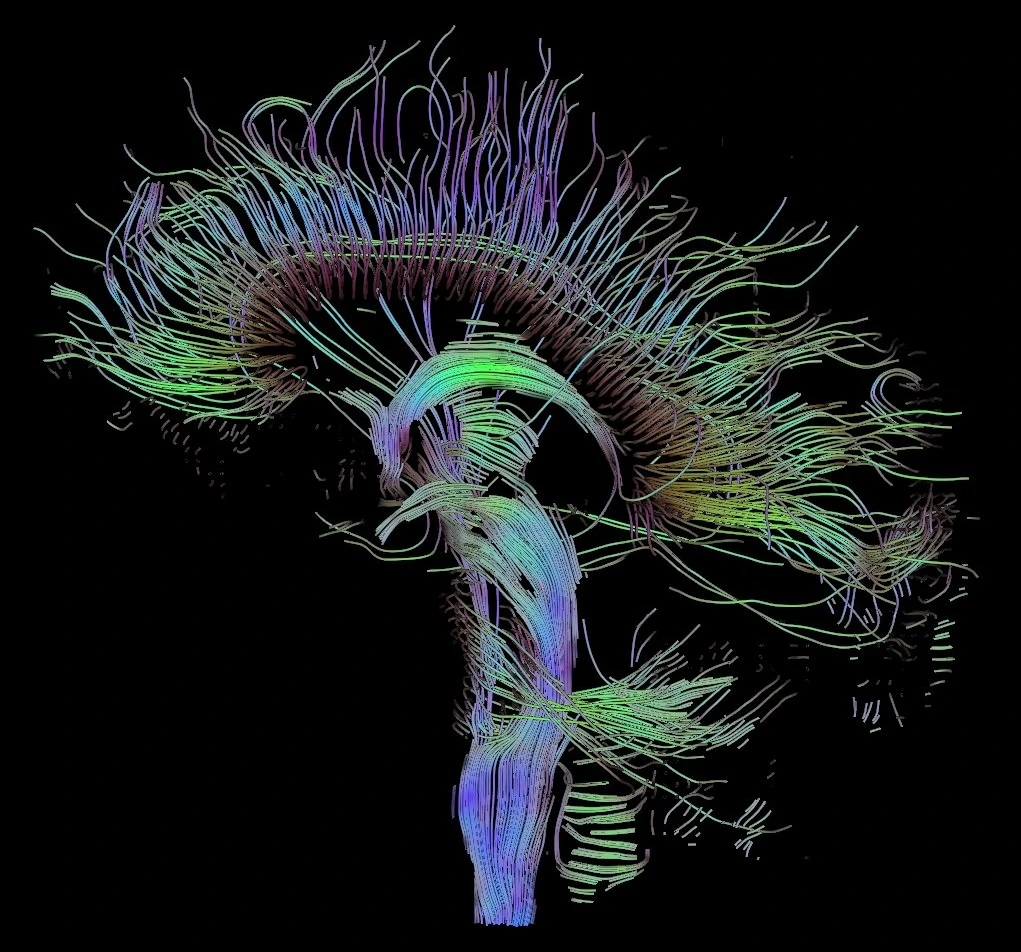
Interpretation
FA describes directional variation among eigenvalues. Crossing or fanning fibers can affect it; FA is not a direct or specific measure of tissue integrity. Inspect the quality of the fitted maps.
Scientific references
Related major models
From the principle to a scoped workflow
A tensor equation does not specify how image volumes, gradient vectors and b-values are paired. Establish their ordering and coordinate frame before interpreting tensor-derived maps.
| Learning path | What to inspect and what it establishes |
|---|---|
| Acquisition, gradient coordinates and bounded fit evidence | Follow the DWI guide for image/gradient alignment, the selected fit, low-b definition, saved map geometry and the limits of the retained sampled comparisons. Evidence scope: The documented comparisons do not establish every estimator, preprocessing route, whole-volume accuracy or clinical interpretation. |
| Keep input geometry and selection with the result | Use the preparation lesson to retain acquisition metadata, data arrangement, identifiers and the actual region or volume supplied to the selected app. Evidence scope: A file extension or successful import alone does not establish gradient orientation or the suitability of a tensor model. |